Common Presentations, corneal issues
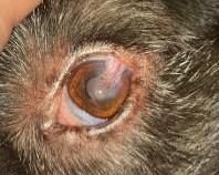
Removal of Corneal Sequestrum
This procedure removes necrotic or damaged tissue from the surface of the cornea to relieve pain and encourage healing.
Depending on the depth and severity of the lesion, additional grafting procedures may be recommended to strengthen the cornea and reduce the risk of rupture.
Some discomfort and discharge are expected during recovery. Healing may take several weeks, and regular follow-up visits are important. Mild scarring or cloudiness can remain after healing, although comfort usually improves significantly after surgery.
Although surgery is generally considered the treatment of choice for this condition, conservative medical management may be offered in selected cases. This approach is discussed individually and requires close monitoring and regular reassessment.
Corneal Diamond Burr Debridement (Diamond Burr Keratectomy)
This treatment removes unhealthy surface cells and anterior stroma from the surface of the cornea and encourages healing.
It is most commonly used for superficial non-healing corneal ulcers (SCCEDs / indolent ulcers).
Mild discomfort and tearing are expected for several days after the procedure. Healing usually occurs within 1-3 weeks, although some patients may require repeat treatment or additional procedures if healing is delayed. In some cases, therapeutic contact lenses may also be used to improve comfort and support healing during recovery.
This treatment can be performed conscious in amenable patients, using local anaesthetic drops applied to numb the eye.
Superficial Keratectomy
This surgery removes abnormal or diseased tissue from the outer layers of the cornea.
It may be recommended for ulceration, pigmentation, inflammatory disease, or certain corneal growths. The aim is to improve comfort, reduce irritation, and preserve vision where possible.
Recovery typically involves eye drops, pain relief, and wearing a protective collar. The eye may appear red, cloudy, or vascular during healing, which is expected to improve over time.
Corneal Graft Surgery (CCT / Biosis / ACell / Amniotic Membrane Grafts and others)
This procedure is used to treat deep corneal ulcers or weakened areas of the cornea that are at risk of rupture. A protective graft is placed over the damaged area to provide structural support, encourage healing, and help preserve the eye and vision. Different graft materials may be used depending on the individual case and severity of disease. Corneal grafts can initially appear red, cloudy, pink, raised, or scarred during healing, which is expected. Recovery requires close monitoring, regular medications, and wearing a protective collar. Although some scarring may remain long-term, these procedures are often essential for saving the eye and significantly improving comfort.Conjunctival Graft
This surgery uses conjunctiva which covers the white of the eye to protect a deep or complicated corneal ulcer and is often performed alongside the use of biomaterials.
The graft provides blood supply and structural support to help the cornea heal safely while reducing the risk of rupture.
Redness, cloudiness, and changes in the appearance of the eye are expected during healing. Although some opacity may remain long-term, the procedure is often very effective at improving comfort and preserving the eye.
UVC Light Therapy
UVC light therapy may be used alongside conventional treatment to help reduce bacterial contamination in corneal infections, particularly infected corneal ulcers.
The treatment is quick, minimally invasive, and usually does not require sedation. It is typically performed in combination with medical therapy.
Corneal Cross-Linking Procedure
Corneal cross-linking is a minimally invasive treatment most commonly used to help stabilise complex ‘melting’ or infected corneal ulcers.
The procedure combines riboflavin eye drops with ultraviolet light to strengthen corneal tissue and improve resistance to further damage.
Cross-linking may help reduce progression of corneal melting and, in some cases, reduce the need for more invasive surgery. Due to the longer treatment time, some patients may require sedation even though the procedure itself is not considered painful.
Foreign Body Removal
Foreign body removal involves removing material trapped in or around the eye that may be causing pain, irritation, or damage.
Foreign material can become lodged under the third eyelid, embedded within the cornea, or in some cases penetrate deeper into the eye. As a result, both the clinical presentation and the degree of ocular damage can vary considerably. Treatment depends on the location and severity of the injury and may range from simple removal under topical anaesthesia to more advanced surgical repair.
After removal, eye drops, pain relief, and close monitoring are often required to ensure proper healing and prevent infection. Prognosis is generally very good when treated promptly, although deeper or penetrating injuries may require more intensive treatment and follow-up care.
It is particularly important not to underestimate penetrating foreign bodies, as these injuries may damage internal eye structures. Of particular concern is damage to the lens as this can result in complications such as cataract formation and severe intraocular inflammation.